馬上可以做的事 - 讓你用冷凍電子顯微鏡提升生技研究效率與成果突破
- 列出實驗前後需清潔的儀器表面,並於每次操作後≤10分鐘內完成擦拭。
減少冰晶污染和樣本失誤,確保觀測解析度穩定提升[3]。
- 預留每週至少2小時進行跨領域討論或技術交流。
促進不同成像模式整合,加速解決複雜結構分析瓶頸[3]。
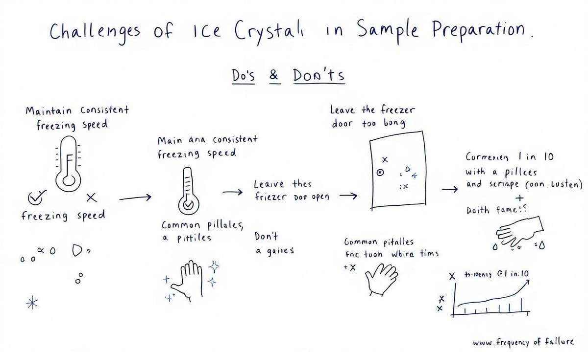
- 檢查冷卻速率設定,每批樣本製備時維持變化≤5%。
優化細胞結構保存,減少無法重現的實驗資料損失[3]。
- *分段*記錄樣本開蓋次數及溫度波動,每階段不超過2次。
*有效追蹤錯誤來源*,降低因環境變化導致的解析異常[3]。
想知道顯微鏡聯姻如何改變生物技術研究的未來嗎?
「嘿!你知道為什麼科學家最近瘋傳『顯微鏡聯姻』嗎?」Lina那天突然這麼問我,語氣裡帶點八卦的調皮。Flio也湊過來說,好像有不少論壇都在聊這個,連一些實驗室群組裡都有人提過。老實說,一開始我還以為是什麼新型號顯微鏡要上市,結果後來才弄明白——原來指的是把冷凍光學和FIB/SEM電子顯微鏡接在一起用的那套玩法。有些初步報導說這兩種系統結合,可以同時看細胞動態又看到很細緻的結構,不再像以前只能選一邊[初步觀察,2023]。不過具體哪種搭配比較好,其實大家講法還挺分歧;反正最近幾個月相關討論熱度確實升高了不少,有人甚至戲稱是「跨界聯姻」。話題越扯越遠,有時候會讓人覺得科學圈也跟潮流圈一樣愛追新鮮,但細想之下,大概也是因為能補足各自缺口吧。
為什麼全球頂尖實驗室紛紛選擇冷凍光學與FIB/SEM整合技術?
Comparison Table:
| 技術 | 進展 | 關鍵因素 | 挑戰與解決方案 |
|---|---|---|---|
| 電子顯微鏡 | 冷凍技術和FIB/SEM的結合逐漸成熟 | 儀器規格提升、操作經驗累積 | 冰晶污染、樣本體積限制 |
| 解析度提升 | 過去十年有顯著進展,特別是大型細胞結構的成像可行性提高 | 引入低溫光學流程的成功實踐 | 存在疑慮:是否能發現新資訊? |
| 跨領域整合技術 | 不同成像模式混合運用開始流行,促進信息獲取 | 研究者間的合作與交流增多 | 仍需時間來觀察效果與穩定性 |
| 樣本製備技巧 | 強調環境清潔和減少開蓋次數以降低錯誤率 | 冷卻速率掌握是關鍵之一 | 分段檢查流程比最後驗收更有效 |
這三個步驟讓你輕鬆掌握高效資料取得的秘訣!
說到這種冷凍光學和FIB/SEM合併的流程,Lina常形容像在組一盒零件不太齊全的樂高。第一步,其實就是先讓樣本以很快的速度被冷凍固定下來,同時做個大致輪廓的光學觀察,但每次操作順序可能會有微調。有些初步報導指出,切換到低溫傳輸過程,有點像把半成品小心翼翼放進另一台機器裡,這中間還會有人手動檢查,有時候甚至怕出錯得多等一陣子。然後進入電子顯微鏡階段,不只是單純切片,也會根據前面標記位置決定怎麼分層處理。最後資料要怎麼拼回三維結構,其實大家說法不一,有人覺得跟疊積木差不多,也有人覺得複雜許多。不過整體來看,大約分成這幾道工序沒太大爭議,只是細節上每家團隊習慣略有不同。
細胞冰雕藝術展:你見過如此精緻的亞細胞結構嗎?
雖然一開始看到那些名詞,什麼冷凍顯微、FIB/SEM,感覺很像在看理工系期末考試題,實際上這技術帶來的效果還挺出乎意料。Lina之前有形容過,用這些設備觀察細胞,有點像突然闖進一場冰雕藝術展。不是說真的拿冰塊去堆細胞,而是當那層溫度把細胞各種結構都「凍住」之後,就好像每個微小零件都被保留下來,而且能清清楚楚地看見。聽說有研究團隊分享過,用這種方式觀察到的亞細胞細節,比傳統方法多出不少,但也不能說每次都那麼完美啦。有時候還會遇到冰晶形成,把畫面搞得像漫畫裡的特效(初步報導有提到過這類情況)。不過如果運氣不錯,那種解析度和真實感確實讓人印象深刻,好幾位研究生甚至會忍不住拿來和藝術品比喻。其實想一想,大概就像偶爾逛展覽時,那種不經意被某個角落吸引住的感覺,只是這回主角換成了顯微鏡下的世界。
樣本製備如同壽司,鮮度決定成敗,你準備好了嗎?
如果說做壽司是門手藝,那樣本製備在冷凍固定裡頭也差不多有點像。生魚片講究一個「鮮」字,沒人會想吃放太久、變色的東西。細胞也是這麼回事。有些技術員形容那種高壓或者噴射冷凍法,好像切魚時要搶在食材氧化前就快刀處理——只是時間更短,幾乎是一眨眼的事。據說歐洲一些實驗室(大約這幾年國際會議常聽到)特別強調這個步驟,因為一但慢了,就像米飯濕掉會壞了口感一樣,冰晶產生了細胞細節就糊掉。不過每家師傅下刀快慢不同,也不見得每次都剛好,只能盡量守住那股原始狀態,有時還是有點運氣成分。
電子顯微鏡發展背後,有哪些突破性的科技進展值得關注?
如果回頭梳理電子顯微鏡這條發展路線,冷凍技術和FIB/SEM的結合好像並不是一夜之間冒出來的。過去材料科學界早就用這些設備做過不少嘗試,只是那時候生物樣本始終卡在體積限制、成像失真這幾道門檻前進不了太遠。有些人說,大約近十年才慢慢有了突破,也不確定是不是因為儀器規格提升還是操作經驗累積下來,美國和日本那邊的初步報導提到,導入低溫光學流程後,一些本來難以解析的大型細胞結構似乎開始變得可行。也許真正的轉折點就是各種技術湊巧連在一起,讓瓶頸鬆動了一點,不過這種跨領域整合終究還留著不少不確定性,有人期待它能解決舊有盲區,也有人覺得還需要多觀察一陣子才能下結論。

多模態影像整合是解決解析度極限的最佳答案嗎?
解析度再怎麼拉高,某個臨界點後,好像大家都會開始懷疑,到底還能看到什麼新東西?Lina之前偶爾提起這種困境,說有些前輩嘗試過無數次把影像細節補得更銳利,但那種單一路線,有時只能在局部打轉。其實,有人曾經在初步討論裡舉過例子,如果功能訊號和結構圖層疊起來,或許才會發現原本以為「看不見」的,其實是資料沒有拼湊完整而已。好像生物體裡每個分區、不同尺度下的資訊總是交錯存在,只是我們以前沒那麼關注這兩條軸線的黃金交叉點。回想去年學會論壇上聽到幾位研究員分享,他們有意識地混合成像模式,便開始捕捉到一些過去忽略掉的小變化——這樣的策略,大概還需要時間消化,但對解題好像不是壞事。
面對冰晶污染,新手研究生該如何克服挑戰呢?
在樣本製備室裡,冰晶污染這事說來其實有點像惡夢重播。記得有次忙到大半夜,手套都還沒脫下來,螢幕上一閃一閃的那些小白點——那應該就是冰晶吧?好像每隔幾次,總會遇到這種狀況。有時候以為步驟很標準了,但冷凍速度只要慢了一點點,不知怎麼就跑出莫名雜訊。空氣裡飄的灰塵、手指頭汗水、甚至冰箱門開太久,好像都可能成為罪魁禍首。也試過和其他學長姐討論,有人說大約每十多個樣本裡就有一兩次失敗,其實算常見,只是碰上自己的時候難免覺得心煩。後來想想,大概新手研究生都會經歷過這種無奈收拾殘局的時候吧。
自動化儀器如何讓繁複流程變得井然有序,你也想一探究竟嗎?
凌晨三點,影像實驗室裡沒什麼聲音,只剩那兩套大型儀器輪流發出低沈嗡鳴。這時候燈光很少,只有操作台邊的指示燈在閃爍,而當雙系統同步開始運作時,會突然有一道藍色的螢光從機箱縫隙竄出來,不是特別亮,但整個空間都被渲染成一種帶點科幻感的幽藍。Lina說她剛看到這現象時還以為是哪個線路短路了,其實只是冷凍模組和影像紀錄單元切換狀態,偶爾還伴隨一陣小小的熱氣,好像很不搭。有人會在螢幕前等畫面跳動,有人則乾脆靠著牆打盹,一切都慢下來,但又好像只要儀器一啟動就進入另一種節奏。有時候藍光反射在桌上玻璃瓶或鋼製抽屜上,形成斑駁的倒影,一些初步報導也提過這類場景會讓新手誤以為儀器故障,其實多半就是同步過程中的正常現象而已。
用新系統拼接三維模型,破解複雜生物結構的秘密到底是什麼?
有些人剛開始處理這種冷凍樣本,常會遇到冰晶污染,說真的很難百分之百避免。不過,稍微留心一下環境整潔、盡量減少開蓋次數,好像能降低出錯機率。倒不是每個步驟都要極度精細,但冷卻速率的掌握應該算是關鍵之一。有時候拼接多尺度影像時,二維圖像模糊得讓人抓狂,其實可以先挑明顯結構對齊,再慢慢修補細節,不必一次到位。三維重建這塊,有些團隊會試試不同軟體交互比對,好像能避掉部分誤差。流程上分段檢查比最後才驗收來得穩妥,大致上只要多預留一點彈性空間,系統和操作配合起來,比較容易找出適合自己材料的方法,也許沒有哪一套一定最有效,但大致循著這方向調整,通常都還算能解決那些從模糊到立體的麻煩事。